As we age, our discs degenerate. Let’s take a look at some MRI images of the spine and different types of degeneration.
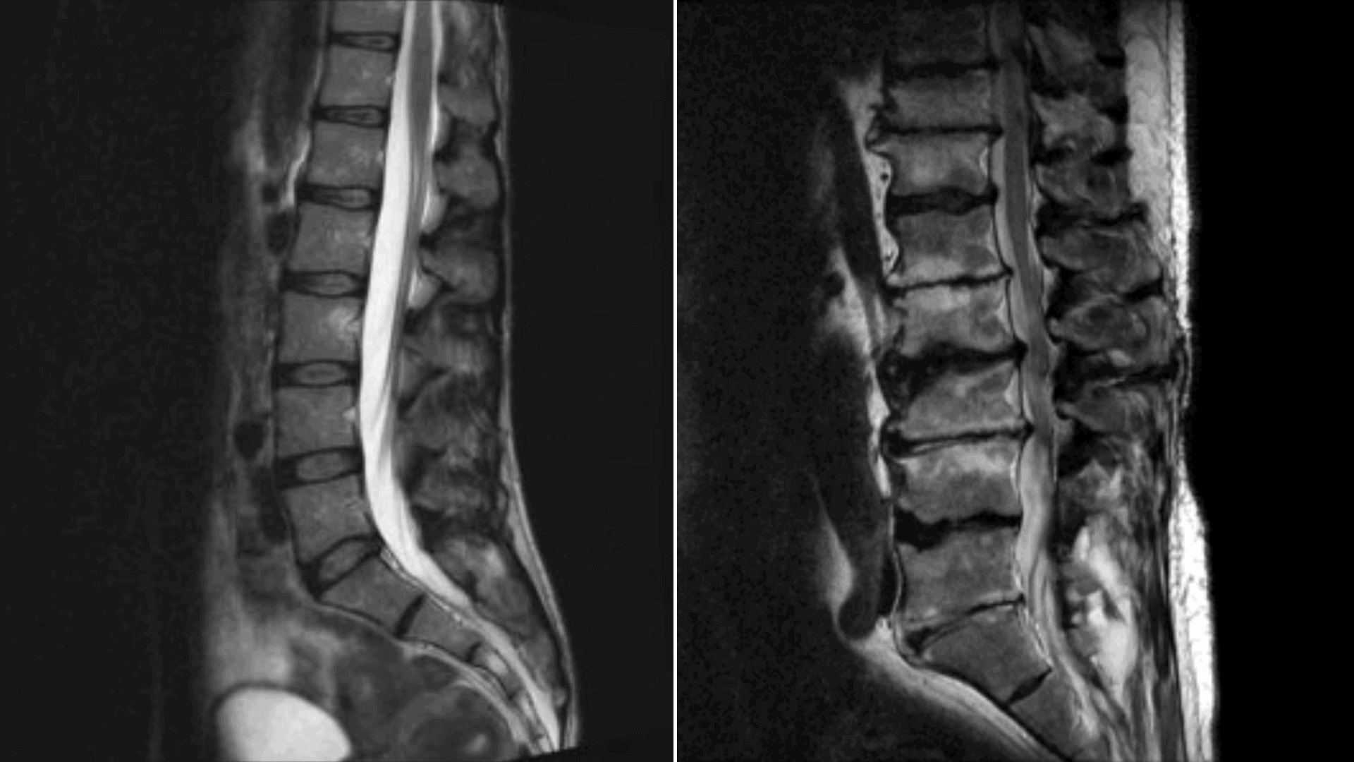
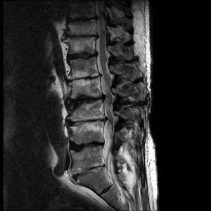
This is a sagittal (side-on) T2 MRI image of a normal lumbar spine in a young adult. As a T2 scan shows water (and fat) as bright, the CSF (spinal fluid)1 is white as is the nucleus2 of the disc. The discs themselves show normal height and no bulging meaning that there is minimal degeneration.
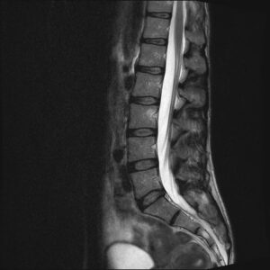
This scan shows degeneration of the lower 4 lumbar discs. The water content is reduced, so the nucleus is darker. The disc height is reduced leading to bulging (similar to when a tyre bulges when some air is let out). There are tiny white specks in the posterior margins of the discs which are annular fissures (degenerative splits).
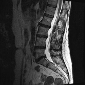
On this scan, there is advanced degeneration of the bottom disc (L5-S1). Compared with the other discs it is very narrow and there is prominent posterior bulging. The bone adjacent to the disc (vertebral endplate) shows increased brightness which is a sign of disc degeneration (Modic changes).
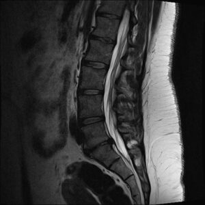
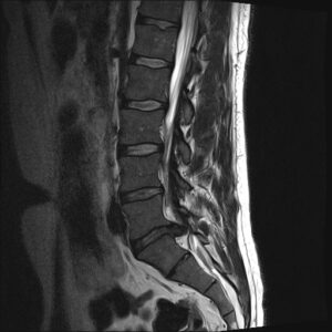 At L5-S1, the back of the disc looks abnormal. This is more than a bulge and is a focal disc herniation. Here, the annulus has split and part of the nucleus has herniated into the spinal canal, compressing the spinal nerves. This type of herniation is called an extrusion. Other types are protrusion and sequestration.
Here, all the discs show advanced degeneration with near-complete loss of height and bulging. The endplates are irregular and show Modic changes. The degeneration has caused the spinal canal to become narrow (stenosis).